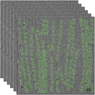
Stack of raw electron micrographs which is used to determine the 3D structure of actin filaments
Link: EMBO J (2020)e104006 https://doi.org/10.15252/embj.2019104006
Actin filaments are one class of cytoskeleton filaments that provide structural framework to cells. The cytoskeleton filament structures are analogous to the scaffolds and columns of a building. However, the cytoskeleton filaments are very dynamic and undergo remodelling in major part of a cell’s life span. Because of their important role in physiology, any deficits or malfunction of actin cytoskeleton and proteins that work along with actin filaments are frequently associated with human diseases. Precise knowledge of the structure of such macromolecules in the cell, is essential for understanding how they function and relevant drug discovery. Recent advances in cryo-electron microscopy (Cryo-EM) has enabled researchers to obtain near-atomic resolution structures of such large macromolecules and understand how mutations affect these structures.
Minhaj Sirajuddin* and his research team at the Cytoskeleton Lab at inStem have determined the structures of actin filament bound to commonly used actin markers. The structures allow for a comparative analysis of three markers bound to actin filament, thus offering valuable insights into their interaction. In Cryo-electron microscopy, protein molecules are frozen in a thin layer of vitreous ice. Interestingly, this specimen for electron microscopy is performed under ‘cryo’ (-180oC) conditions and hence the technique is popularly called cryo-EM. When this sample is subjected to a beam of electrons, a 2D image projection of embedded proteins is created. Because the protein molecules in the vitreous ice can randomly orient in many different ways, the 2D images are back projected in Fourier space to create a 3D shape of the protein molecules. Filaments like actins have repeating arrays of same molecule and thus 3D structures of such filaments can be obtained with cryo-EM and helical reconstruction. The National Cryo-EM Facility on campus, funded by DBT has enabled Minhaj’s team to analyse cryo-EM structures of actin filament bound to actin markers.
Fluorescent markers that specifically label cellular structures can be visualized using a fluorescent microscopy. This is widely used in cell biology to study molecules inside a cellular environment. Thus, cellular studies of filamentous actin (F-actin) process commonly utilize fluorescent versions of toxins, peptides, and proteins that bind actin. While the choice of these markers has been largely based on availability and ease, there is a severe dearth of structural data for an informed judgment in employing suitable F-actin markers for a particular requirement. In the current research work, the team described the cryo-EM structures of phalloidin (toxin), lifeAct (peptide) and utrophin (actin-binding protein) bound to F-actin, providing a comprehensive high-resolution structural comparison of widely used actin markers and their influence towards F-actin. Cryo-EM structures, biochemical and cell biological analysis of these three probes bound to actin filaments offer guidance for their respective suitability:
-
Phalloidin binding to actin filament does not induce actin conformational changes.
-
LifeAct preferentially binds to the closed D-loop conformation of actin filaments.
-
The utrophin CH1 domain is sufficient for interaction with F-actin.
Actin filaments have been studied for many decades now and researchers have understood its intriguing role in cell movement, muscle contraction, and maintaining cell shape and its integrity. While there are many reports on suitability of these fluorescent actin markers, none account for how these actin markers interact with actin filament. “The current work will aid researchers in choosing and designing better probes with desirable qualities to label cellular actin in the future,” says Minhaj. Further, actin filament and associated protein molecules are frequently associated with mutations that cause cardiomyopathies. Minhaj’s work with actin filaments bound to cellular markers has provided a pipeline to explore cryo-EM structure determination methods in studying actin and related gene products associated with cardiomyopathies. We are hopeful that such research findings will help in better understanding of cardiac diseases.
*Minhaj Sirajuddin’s laboratory at the Centre for Cardiovascular Disorders (CCBD) at inStem, has exploited the power of cryo-EM (Cryogenic Electron Microscopy), to determine the structures of actin filament bound to commonly used actin markers (Kumari A., Kesarwani S., Javoor MG., Vinothkumar KR & Sirajuddin M. EMBO J (2020)e104006 https://doi.org/10.15252/embj.2019104006). This work is a collaboration with Vinothkumar K Ragunath, NCBS. Minhaj Sirajuddin is an EMBO YIP Investigator and a recipient of a Wellcome Trust- DBT India Alliance Intermediate Fellowship.
Reference:
Structural insights into actin filament recognition by commonly used cellular actin markers
Archana Kumari, Shubham Kesarwani, Manjunath G Javoor, Kutti R Vinothkumar, and Minhajuddin Sirajuddin, June 2020, The EMBO Journal
https://www.embopress.org/doi/full/10.15252/embj.2019104006
Publication Date: June 22, 2020
Media Coverage: